포커스
포커스Ⅱ
Here comes the SUN: 2021 SUN updates
한국포도막학회 편집위원회
1969년 발표한 비틀즈의 노래 ‘Here comes the sun’을 들으면 감미로운 어쿠스틱 기타 선율, 그리고 그 당시에는 새로운 시도였던 신디사이저와의 조화가 지금 들어도 전혀 촌스럽지 않고 반세기를 지난 지금도 청중들의 귀를 즐겁게 합니다. 고전은 동서 고금을 막론하고 통(?)한다는 말을 실감케 하는데, 포도막염 환자를 보는 임상 의사에게 SUN classification은 출판된지 16년이 지난
어찌보면 고전이면서도, 매일 따뜻하게 빛을 비춰주는 해처럼 우리의 일상 진료에 영향을 미치는 것이 아닐까 싶습니다.
그런 SUN classification이 최근 American Journal of Ophthalmology에 많은 숫자의 리포트를 통해 업데이트 되었습니다. 글 제목과 똑같은 이름의 editorial1 과 함께 26개의 disease classification criteria에 관한 논문을 발표했는데, 갑작스런 정 보의 홍수가 아찔하기는 하지만 핵심적인 내용들을 중심으로 지면을 통해 소개드리고자 합니다.
SUN classification update의 대략적인 구성은 ‘1+25’로 요약할 수 있습니다. 우선 전체 포도막염 질환들의 분류에 대해 다루는 총론격인 논문이 있고,2 여기서 제시한 총 25가지 세부 질환 각각에 대한 분류를 다루는 각론에 해당하는 25편의 논문들이 있습니다.3-27
우선 SUN working group에서 약 10년에 걸친 이 대형 프로젝트를 위해 사용한 방법론 은 크게 4단계로 나눠볼 수 있는데
① informatics – 질환 및 증례 기술을 위한 용어의 표준화 및 증례 수집을 위한 시스템 개발
② case collection – 25개 흔한 포도막염 질환 5,766 케이스를 76명의 참여 연구자로 부터 받아 각종 검사 영상들과 함께 데이터베이스에 축적(각 질환마다 약 250 증례가 될 때까지 수집)하고 reading center 에서 영상들로부터 병변 크기, 수 등 특징을 추출
③ case selection – 특정 질환의 진단 기준을 정하기 위해 전문가로부터 ‘supermajority(>75%) agreement’를 보인 케이스를 선별하여 최종 데이터베이스에 보관
④ machine learning – 증례들을 training set, validation set 으로 나누고, misclassification을 최소화할 수 있는 진단 기준을 machine learning을 통해 개발하여 validation set 에서 검증하는 단계입니다.
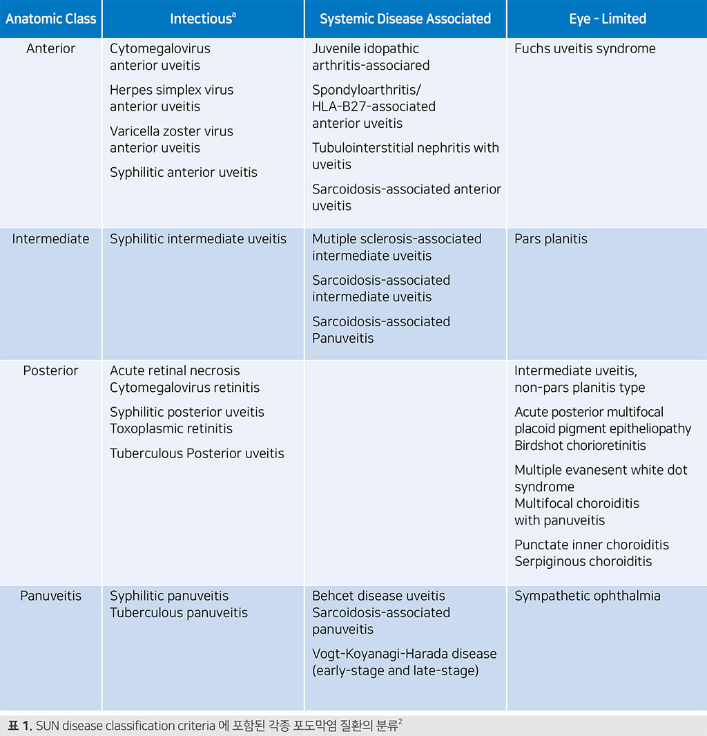
2021 SUN classification 은 각종 포도막염 질환의 분류를 두 가지 축으로 분류하였 는데, 한가지는 해부학적 위치(anterior, intermediate, posterior, panuveitis), 다른 하나는 병인(infectious, systemic disease associated, eye-limited) 입니다. 여기서 systemic disease는 주로 전신 자가면역질환을 일컫고, eye-limited는 immune-mediated 기전으로 생각되는, 눈에 국한된 질환들입니다. 이를 두 축으로 SUN에서 제시하는 포도막염 질환의 분류는 다음 표(표 1)와 같습니다.
| Anatomic Class | Intectiousa | Systemic Disease Associated | Eye - Limited | ||
|---|---|---|---|---|---|
| Anterior |
Cytomegalovirus anterior uveitis Herpes simplex virus anterior uveitis Varicella zoster virus anterior uveitis Syphilitic anterior uveitis |
Juvenile idopathic arthritis-associared Spondyloarthritis/HLA-B27-associated anterior uveitis Tubulointerstitial nephritis with uveitis Sarcoidosis-associated anterior uveitis |
Fuchs uveitis syndrome |
||
| Intermediate |
Syphilitic intermediate uveitis |
Mutiple sclerosis-associated intermediate uveitis Sarcoidosis-associated intermediate uveitis Sarcoidosis-associated Panuveitis |
Pars planitis |
||
| Posterior |
Acute retinal necrosis Cytomegalovirus retinitis Syphilitic posterior uveitis Toxoplasmic retinitis Tuberculous Posterior uveitis |
Intermediate uveitis, non-pars planitis type Acute posterior multifocal placoid pigment epitheliopathy Birdshot chorioretinitis Multiple evanesent white dot syndrome Multifocal choroiditis with panuveitis Punctate inner choroiditis Serpiginous choroiditis |
|||
| Panuveitis |
Syphilitic panuveitis Tuberculous panuveitis |
Behcet disease uveitis Sarcoidosis-associated panuveitis Vogt-Koyanagi-Harada disease |
Sympathetic ophthalmia |
||
| 표 1. SUN disease classification criteria 에 포함된 각종 포도막염 질환의 분류 2 | |||||
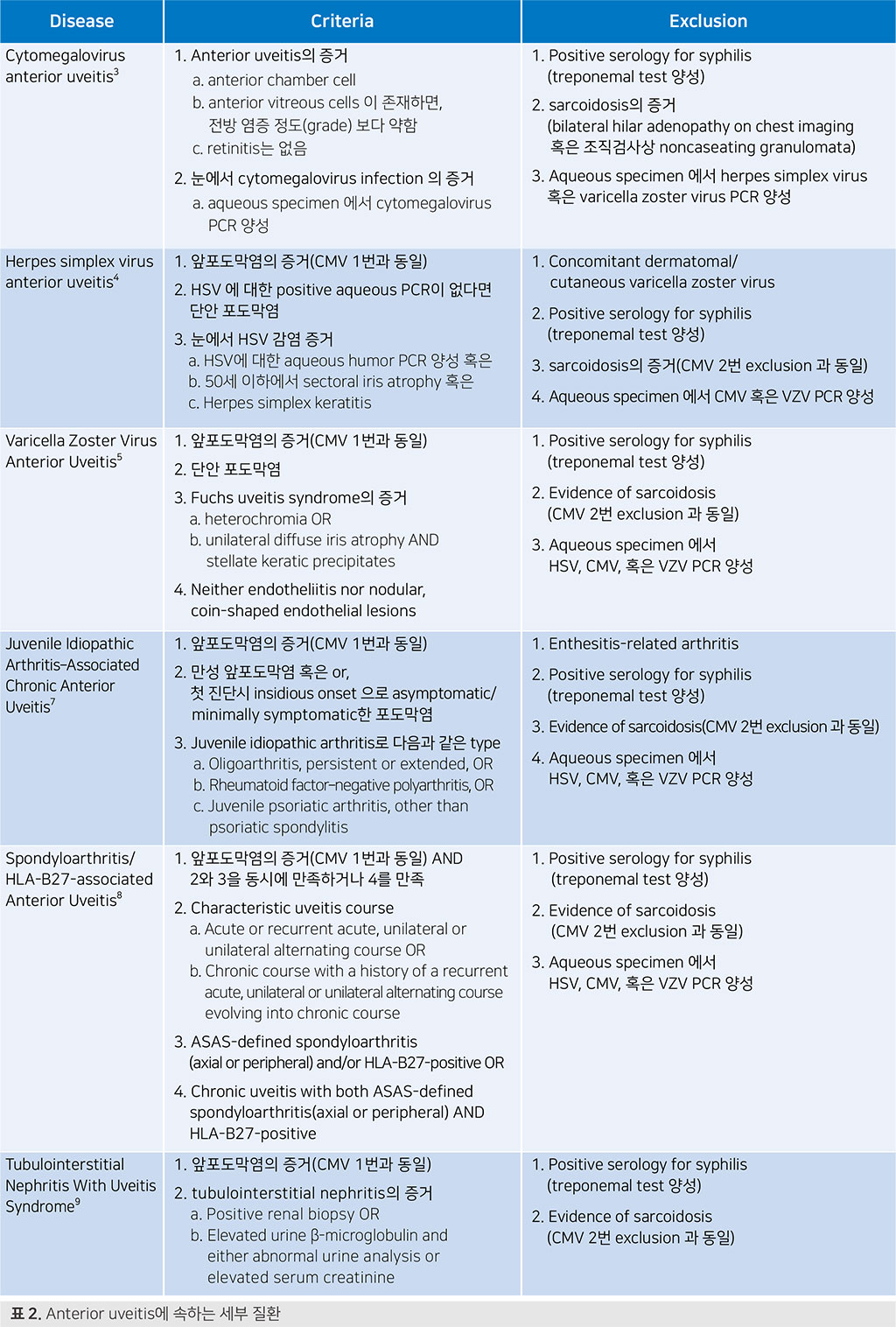
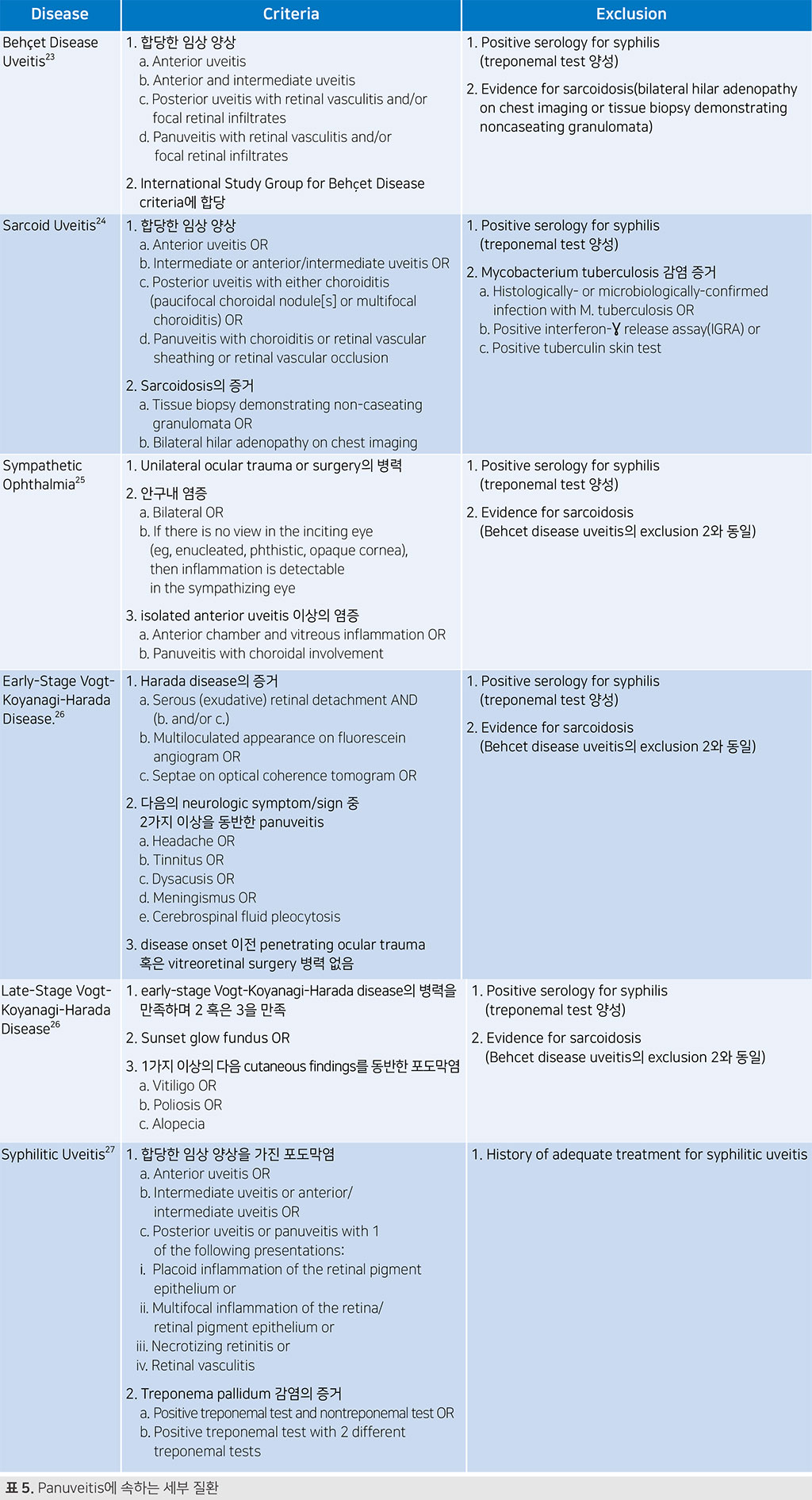
그럼 해부학적 분류(anterior, intermediate, posterior, panuveitis)에 따라 각 분류에 속하는 세부질환들의 진단 및 배제 기준을 살펴보겠습니다.
| Disease | Criteria | Exclusion | |||
|---|---|---|---|---|---|
| Cytomegalovirus anterior uveitis 3 |
1. Anterior uveitis의 증거 a. anterior chamber cell b. anterior vitreous cells 이 존재하면, 전방 염증 정도(grade) 보다 약함 c. retinitis는 없음 2. 눈에서 cytomegalovirus infection 의 증거 a. aqueous specimen 에서 cytomegalovirus PCR 양성 |
1. Positive serology for syphilis(treponemal test 양성) 2. sarcoidosis의 증거 (bilateral hilar adenopathy on chest imaging 혹은 조직검사상 noncaseating granulomata) 3. Aqueous specimen 에서 herpes simplex virus 혹은 varicella zoster virus PCR 양성 |
|||
| Herpes simplex virus anterior uveitis 4 |
1. 앞포도막염의 증거(CMV 1번과 동일) 2. HSV 에 대한 positive aqueous PCR 이 없다면 단안 포도막염 3. 눈에서 HSV 감염 증거 a. HSV에 대한 aqueous humor PCR 양성 혹은 b. 50세 이하에서 sectoral iris atrophy 혹은 c. Herpes simplex keratitis |
1. Concomitant dermatomal/cutaneous varicella zoster virus 2. Positive serology for syphilis(treponemal test 양성) 3. sarcoidosis의 증거(CMV 2번 exclusion 과 동일) 4. Aqueous specimen 에서 CMV 혹은 VZV PCR 양성 |
|||
| Varicella Zoster Virus Anterior Uveitis 5 |
1. 앞포도막염의 증거(CMV 1번과 동일) 2. 단안 포도막염 3. Fuchs uveitis syndrome의 증거 a. heterochromia OR b. unilateral diffuse iris atrophy AND stellate keratic precipitates 4. Neither endotheliitis nor nodular, coin-shaped endothelial lesions |
1. Positive serology for syphilis(treponemal test 양성) 2. Evidence of sarcoidosis(CMV 2번 exclusion 과 동일) 3. Aqueous specimen 에서 HSV, CMV, 혹은 VZV PCR 양성 |
|||
| Juvenile Idiopathic Arthritis–Associated Chronic Anterior Uveitis 7 |
1. 앞포도막염의 증거(CMV 1번과 동일) 2. 만성 앞포도막염 혹은 or, 첫 진단시 insidious onset 으로 asymptomatic/minimally symptomatic 한 포도막염 3. Juvenile idiopathic arthritis로 다음과 같은 type a. Oligoarthritis, persistent or extended, OR b. Rheumatoid factor–negative polyarthritis, OR c. Juvenile psoriatic arthritis, other than psoriatic spondylitis |
1. Enthesitis-related arthritis 2. Positive serology for syphilis(treponemal test 양성) 3. Evidence of sarcoidosis(CMV 2번 exclusion 과 동일) 4. Aqueous specimen 에서 HSV, CMV, 혹은 VZV PCR 양성 |
|||
| Spondyloarthritis/ HLA-B27-associated Anterior Uveitis 8 |
1. 앞포도막염의 증거(CMV 1번과 동일) AND 2와 3을 동시에 만족하거나 4를 만족 2. Characteristic uveitis course a. Acute or recurrent acute, unilateral or unilateral alternating course OR b. Chronic course with a history of a recurrent acute, unilateral or unilateral alternating course evolving into chronic course 3. ASAS-defined spondyloarthritis (axial or peripheral) and/or HLA-B27-positive OR 4. Chronic uveitis with both ASAS-defined spondyloarthritis (axial or peripheral) AND HLA-B27-positive |
1. Positive serology for syphilis(treponemal test 양성) 2. Evidence of sarcoidosis(CMV 2번 exclusion 과 동일) 3. Aqueous specimen 에서 HSV, CMV, 혹은 VZV PCR 양성 |
|||
| Tubulointerstitial Nephritis With Uveitis Syndrome 9 |
1. 앞포도막염의 증거(CMV 1번과 동일) 2. tubulointerstitial nephritis의 증거 a. Positive renal biopsy OR b. Elevated urine β-microglobulin and either abnormal urine analysis or elevated serum creatinine |
1. Positive serology for syphilis(treponemal test 양성) 2. Evidence of sarcoidosis(CMV 2번 exclusion 과 동일) |
|||
| 표 2. Anterior uveitis에 속하는 세부 질환 | |||||
| Disease | Criteria | Exclusion | |||
|---|---|---|---|---|---|
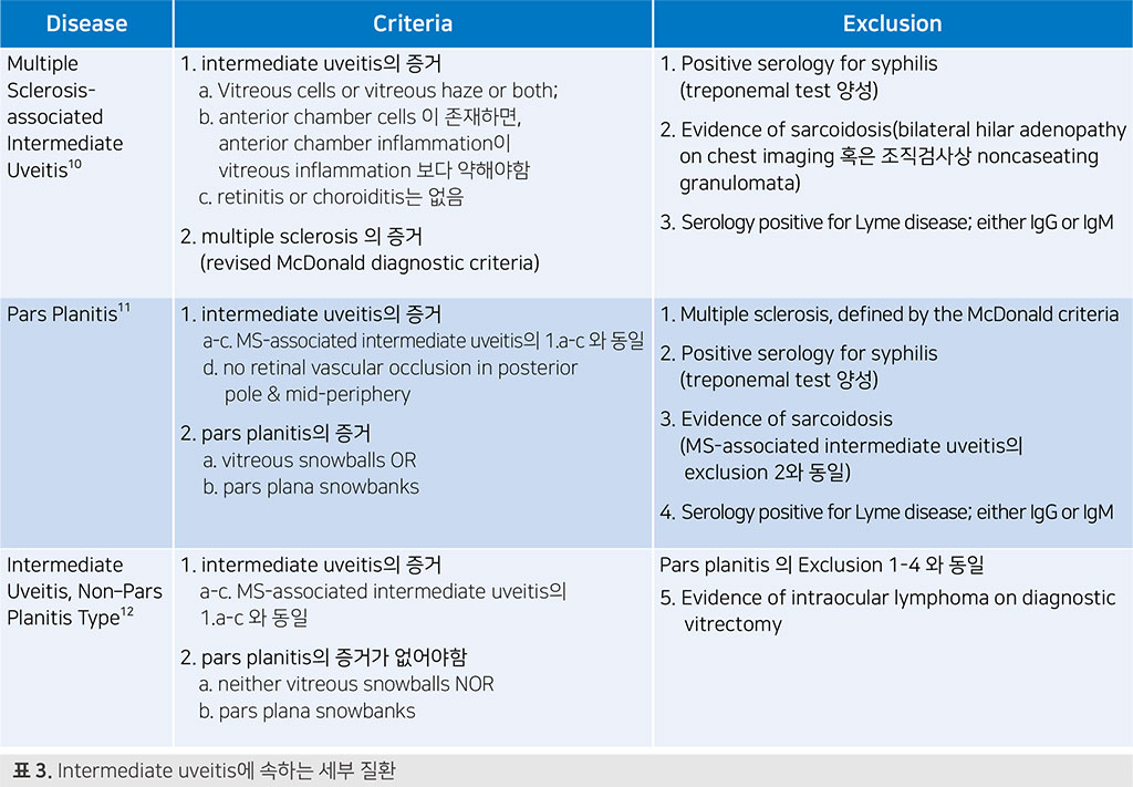
| Multiple Sclerosisassociated Intermediate Uveitis 10 |
1. intermediate uveitis의 증거 a. Vitreous cells or vitreous haze or both; b. anterior chamber cells 이 존재하면, anterior chamber inflammation이 vitreous inflammation 보다 약해야함 c. retinitis or choroiditis는 없음
2. multiple sclerosis 의 증거 |
1. Positive serology for syphilis(treponemal test 양성) 2. Evidence of sarcoidosis(bilateral hilar adenopathy on chest imaging 혹은 조직검사상 noncaseating granulomata) 3. Serology positive for Lyme disease; either IgG or IgM |
|||
| Pars Planitis 11 |
1. intermediate uveitis의 증거 a-c. MS-associated intermediate uveitis 의 1.a-c 와 동일 d. no retinal vascular occlusion in posterior pole & mid-periphery 2. pars planitis의 증거 a. vitreous snowballs OR b. pars plana snowbanks |
1. Multiple sclerosis, defined by the McDonald criteria 2. Positive serology for syphilis(treponemal test 양성) 3. Evidence of sarcoidosis(MS-associated intermediate uveitis 의 exclusion 2와 동일) 4. Serology positive for Lyme disease; either IgG or IgM |
|||
| Intermediate Uveitis, Non–Pars Planitis Type 12 |
1. intermediate uveitis의 증거 a-c. MS-associated intermediate uveitis 의 1.a-c 와 동일 2. pars planitis의 증거가 없어야함 a. neither vitreous snowballs NOR b. pars plana snowbanks |
Pars planitis 의 Exclusion 1-4 와 동일 5. Evidence of intraocular lymphoma on diagnostic vitrectomy |
|||
| 표 3. Intermediate uveitis에 속하는 세부 질환 | |||||
| Disease | Criteria | Exclusion | |||
|---|---|---|---|---|---|
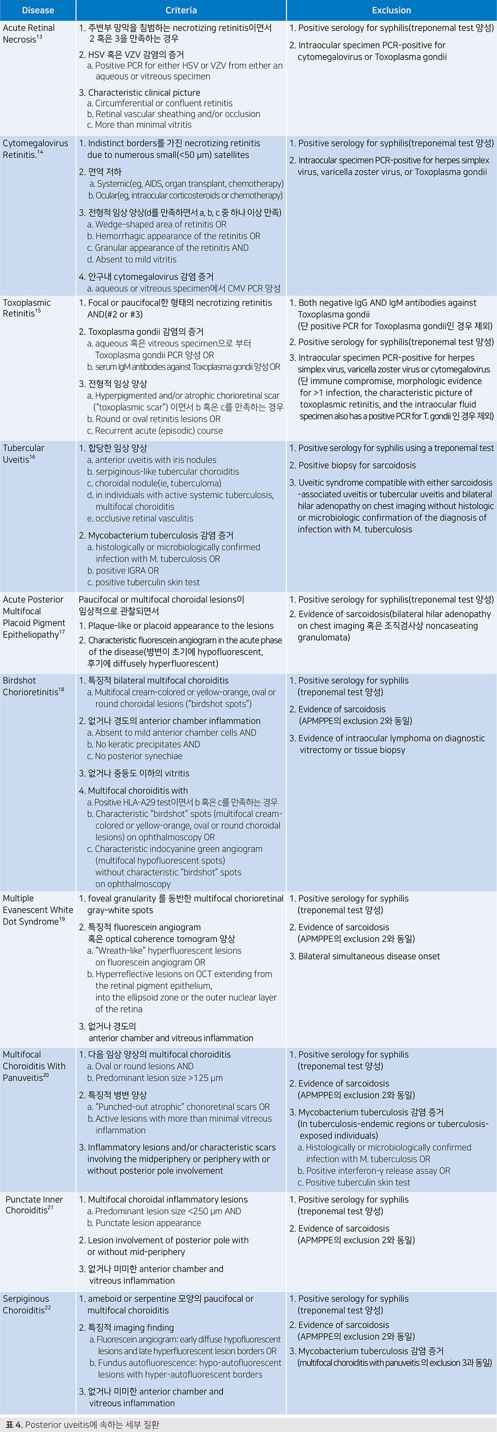
| Acute Retinal Necrosis 13 |
1. 주변부 망막을 침범하는 necrotizing retinitis이면서 2 혹은 3을 만족하는 경우 2. HSV 혹은 VZV 감염의 증거 a. Positive PCR for either HSV or VZV from either an aqueous or vitreous specimen 3. Characteristic clinical picture a. Circumferential or confluent retinitis b. Retinal vascular sheathing and/or occlusion c. More than minimal vitritis |
1. Positive serology for syphilis(treponemal test 양성) 2. Intraocular specimen PCR-positive for cytomegalovirus or Toxoplasma gondii |
|||
| Cytomegalovirus Retinitis. 14 |
1. Indistinct borders를 가진 necrotizing retinitis due to numerous small(<50 μm) satellites 2. 면역 저하 a. Systemic (eg, AIDS, organ transplant, chemotherapy) b. Ocular (eg, intraocular corticosteroids or chemotherapy) 3. 전형적 임상 양상(d를 만족하면서 a, b, c 중 하나 이상 만족) a. Wedge-shaped area of retinitis OR b. Hemorrhagic appearance of the retinitis OR c. Granular appearance of the retinitis AND d. Absent to mild vitritis |
1. Positive serology for syphilis(treponemal test 양성) 2. Intraocular specimen PCR-positive for herpes simplex virus, varicella zoster virus, or Toxoplasma gondii |
|||
| Toxoplasmic Retinitis 15 |
1. Focal or paucifocal한 형태의 necrotizing retinitis AND(#2 or #3) 2. Toxoplasma gondii 감염의 증거 a. aqueous 혹은 vitreous specimen으로 부터 Toxoplasma gondii PCR 양성 OR b. serum IgM antibodies against Toxoplasma gondii 양성 OR 3. 전형적 임상 양상 a. Hyperpigmented and/or atrophic chorioretinal scar (“toxoplasmic scar”) 이면서 b 혹은 c를 만족하는 경우 b. Round or oval retinitis lesions OR c. Recurrent acute (episodic) course |
1. Both negative IgG AND IgM antibodies against Toxoplasma gondii (단 positive PCR for Toxoplasma gondii인 경우 제외) 2. Positive serology for syphilis(treponemal test 양성) 3. Intraocular specimen PCR-positive for herpes simplex virus, varicella zoster virus or cytomegalovirus (단 immune compromise, morphologic evidence for >1 infection, the characteristic picture of toxoplasmic retinitis, and the intraocular fluid specimen also has a positive PCR for T. gondii 인 경우 제외) |
|||
| Tubercular Uveitis 16 |
1. 합당한 임상 양상 a. anterior uveitis with iris nodules b. serpiginous-like tubercular choroiditis c. choroidal nodule(ie, tuberculoma) d. in individuals with active systemic tuberculosis, multifocal choroiditis e. occlusive retinal vasculitis 2. Mycobacterium tuberculosis 감염 증거 a. histologically or microbiologically confirmed infection with M. tuberculosis OR b. positive IGRA OR c. positive tuberculin skin test |
1. Positive serology for syphilis using a treponemal test 2. Positive biopsy for sarcoidosis 3. Uveitic syndrome compatible with either sarcoidosis -associated uveitis or tubercular uveitis and bilateral hilar adenopathy on chest imaging without histologic or microbiologic confirmation of the diagnosis of infection with M. tuberculosis |
|||
| Acute Posterior Multifocal Placoid Pigment Epitheliopathy 17 |
Paucifocal or multifocal choroidal lesions 이임상적으로 관찰되면서 1. Plaque-like or placoid appearance to the lesions 2. Characteristic fluorescein angiogram in the acute phase of the disease(병변이 초기에 hypofluorescent, 후기에 diffusely hyperfluorescent) |
1. Positive serology for syphilis(treponemal test 양성) 2. Evidence of sarcoidosis(bilateral hilar adenopathy on chest imaging 혹은 조직검사상 noncaseating granulomata) |
|||
| Birdshot Chorioretinitis 18 |
1. 특징적 bilateral multifocal choroiditis a. Multifocal cream-colored or yellow-orange, oval or round choroidal lesions (“birdshot spots”) 2. 없거나 경도의 anterior chamber inflammation a. Absent to mild anterior chamber cells AND b. No keratic precipitates AND c. No posterior synechiae 3. 없거나 중등도 이하의 vitritis 4. Multifocal choroiditis with a. Positive HLA-A29 test이면서 b 혹은 c를 만족하는 경우 b. Characteristic “birdshot” spots (multifocal creamcolored or yellow-orange, oval or round choroidal lesions) on ophthalmoscopy OR c. Characteristic indocyanine green angiogram (multifocal hypofluorescent spots) without characteristic “birdshot” spots on ophthalmoscopy |
1. Positive serology for syphilis(treponemal test 양성) 2. Evidence of sarcoidosis(APMPPE의 exclusion 2와 동일) 3. Evidence of intraocular lymphoma on diagnostic vitrectomy or tissue biopsy |
|||
| Multiple Evanescent White Dot Syndrome 19 |
1. foveal granularity 를 동반한 multifocal chorioretinal gray-white spots 2. 특징적 fluorescein angiogram 혹은 optical coherence tomogram 양상 a. “Wreath-like” hyperfluorescent lesions on fluorescein angiogram OR b. Hyperreflective lesions on OCT extending from the retinal pigment epithelium, into the ellipsoid zone or the outer nuclear layer of the retina 3. 없거나 경도의 anterior chamber and vitreous inflammation |
1. Positive serology for syphilis(treponemal test 양성) 2. Evidence of sarcoidosis(APMPPE의 exclusion 2와 동일) 3. Bilateral simultaneous disease onset |
|||
| Multifocal Choroiditis With Panuveitis 20 |
1. 다음 임상 양상의 multifocal choroiditis a. Oval or round lesions AND b. Predominant lesion size >125 μm 2. 특징적 병변 양상 a. “Punched-out atrophic” chorioretinal scars OR b. Active lesions with more than minimal vitreous inflammation 3. Inflammatory lesions and/or characteristic scars involving the midperiphery or periphery with or without posterior pole involvement |
1. Positive serology for syphilis(treponemal test 양성) 2. Evidence of sarcoidosis(APMPPE의 exclusion 2와 동일) 3. Mycobacterium tuberculosis 감염 증거(In tuberculosisendemic regions or tuberculosis-exposed individuals) a. Histologically or microbiologically confirmed infection with M. tuberculosis OR b. Positive interferon-γ release assay OR c. Positive tuberculin skin test |
|||
| Punctate Inner Choroiditis 21 |
1. Multifocal choroidal inflammatory lesions a. Predominant lesion size <250 μm AND b. Punctate lesion appearance 2. Lesion involvement of posterior pole with or without mid-periphery 3. 없거나 미미한 anterior chamber and vitreous inflammation |
1. Positive serology for syphilis(treponemal test 양성) 2. Evidence of sarcoidosis(APMPPE의 exclusion 2와 동일) |
|||
| Serpiginous Choroiditis 22 |
1. ameboid or serpentine 모양의 paucifocal or multifocal choroiditis 2. 특징적 imaging finding a. Fluorescein angiogram: early diffuse hypofluorescent lesions and late hyperfluorescent lesion borders OR b. Fundus autofluorescence: hypo-autofluorescent lesions with hyper-autofluorescent borders 3. 없거나 미미한 anterior chamber and vitreous inflammation |
1. Positive serology for syphilis(treponemal test 양성) 2. Evidence of sarcoidosis(APMPPE의 exclusion 2와 동일) 3. Mycobacterium tuberculosis 감염 증거 (multifocal choroiditis with panuveitis 의 exclusion 3과 동일) |
|||
| 표 4. Posterior uveitis에 속하는 세부 질환 | |||||
| Disease | Criteria | Exclusion | |||
|---|---|---|---|---|---|
| Behçet Disease Uveitis 23 |
1. 합당한 임상 양상 a. Anterior uveitis b. Anterior and intermediate uveitis c. Posterior uveitis with retinal vasculitis and/or focal retinal infiltrates d. Panuveitis with retinal vasculitis and/or focal retinal infiltrates 2. International Study Group for Behçet Disease criteria에 합당 |
1. Positive serology for syphilis(treponemal test 양성) 2. Evidence for sarcoidosis(bilateral hilar adenopathy on chest imaging or tissue biopsy demonstrating noncaseating granulomata) |
|||
| Sarcoid Uveitis 24 |
1. 합당한 임상 양상 a. Anterior uveitis OR b. Intermediate or anterior/intermediate uveitis OR c. Posterior uveitis with either choroiditis (paucifocal choroidal nodule[s] or multifocal choroiditis) OR d. Panuveitis with choroiditis or retinal vascular sheathing or retinal vascular occlusion 2. Sarcoidosis의 증거 a. Tissue biopsy demonstrating non-caseating granulomata OR b. Bilateral hilar adenopathy on chest imaging |
1. Positive serology for syphilis(treponemal test 양성) 2. Mycobacterium tuberculosis 감염 증거 a. Histologically- or microbiologically-confirmed infection with M. tuberculosis OR b. Positive interferon-Ɣ release assay(IGRA) or c. Positive tuberculin skin test |
|||
| Sympathetic Ophthalmia 25 |
1. Unilateral ocular trauma or surgery의 병력 2. 안구내 염증 a. Bilateral OR b. If there is no view in the inciting eye (eg, enucleated, phthistic, opaque cornea), then inflammation is detectable in the sympathizing eye 3. isolated anterior uveitis 이상의 염증 a. Anterior chamber and vitreous inflammation OR b. Panuveitis with choroidal involvement |
1. Positive serology for syphilis(treponemal test 양성) 2. Evidence for sarcoidosis (Behcet disease uveitis의 exclusion 2와 동일) |
|||
| Early-Stage VogtKoyanagi-Harada Disease. 26 |
1. Harada disease의 증거 a. Serous (exudative) retinal detachment AND(b. and/or c.) b. Multiloculated appearance on fluorescein angiogram OR c. Septae on optical coherence tomogram OR 2. 다음의 neurologic symptom/sign 중 2가지 이상을 동반한 panuveitis a. Headache OR b. Tinnitus OR c. Dysacusis OR d. Meningismus OR e. Cerebrospinal fluid pleocytosis 3. disease onset 이전 penetrating ocular trauma 혹은 vitreoretinal surgery 병력 없음 |
1. Positive serology for syphilis(treponemal test 양성) 2. Evidence for sarcoidosis (Behcet disease uveitis의 exclusion 2와 동일) |
|||
| Late-Stage VogtKoyanagi-Harada Disease 26 |
1. early-stage Vogt-Koyanagi-Harada disease의 병력을 만족하며 2 혹은 3을 만족 2. Sunset glow fundus OR 3. 1가지 이상의 다음 cutaneous findings를 동반한 포도막염 a. Vitiligo OR b. Poliosis OR c. Alopecia |
1. Positive serology for syphilis(treponemal test 양성) 2. Evidence for sarcoidosis (Behcet disease uveitis의 exclusion 2와 동일) |
|||
| Syphilitic Uveitis 27 |
1. 합당한 임상 양상을 가진 포도막염 a. Anterior uveitis OR b. Intermediate uveitis or anterior/intermediate uveitis OR c. Posterior uveitis or panuveitis with 1 of the following presentations: i. Placoid inflammation of the retinal pigment epithelium or ii. Multifocal inflammation of the retina/retinal pigment epithelium or iii. Necrotizing retinitis or iv. Retinal vasculitis 2. Treponema pallidum 감염의 증거 a. Positive treponemal test and nontreponemal test OR b. Positive treponemal test with 2 different treponemal tests |
1. History of adequate treatment for syphilitic uveitis |
|||
| 표 5. Panuveitis에 속하는 세부 질환 | |||||
10년간에 걸쳐 많은 케이스를 수집하고 여러 단계에 걸친 체계적인 작업들을 수행하여 수십편의 연구 논문으로 완성한 매우 훌륭하고 의미있는 프로젝트 같습니다. 어찌보면 포 도막염이 양상이 매우 다양하고 consensus가 필요한 질환이기 때문에 이런 작업이 절실 한데, 이번 update는 첫 SUN classification이 그랬듯 앞으로 임상적으로나 연구 차원에 서도 큰 영향력이 있는 classification이라 생각됩니다. 선별 작업을 거친 좋은 증례들을 바탕으로 최소한의 오류를 위해 classification criteria를 만들 때 machine learning을 사용하였다는 것이 실험적이면서도 참신한 아이디어 같습니다. 앞으로의 의학에서 질병 분류에 관해 SUN classification은 방법론적으로도 모범적 예가 될 것으로 생각됩니다.
한편으로 2005년 SUN classification 과 이번 update를 보면서, 그 사이 의학의 방법론에 있어 informatics를 이용하며, consensus 를 이루는 많은 방법론의 발전과 함께 격 세지감을 느낄 수 있었습니다. 투명화, 정보화, 인공지능화? 라고나 표현해야 할까요, 포 도막염은 몇몇 대가 선생님들께 직접 환자를 진료하고 치료하시는걸 보면서 그 진단 및 치료 철학을 통해 배우는 분야라 생각했는데, 점점 소수의 영향력 보다는 잘 만들어진 시스템으로 consensus가 만들어지고 움직이는 것 같다는 생각이 들었습니다. 학회를 중심으로 포도막염에 관심있으신 회원님들의 활발한 협력, 공동 연구를 고대하며, 또 무엇보다 조금이나마 본 글을 통해 회원님들의 포도막염 진료와 연구에 도움되시길 소망하며 글을 마칩니다.
- 1 Van Gelder RN, Sen N, Tufail A, Lee AY. Here comes the SUN (Part 2): Standardization of uveitis nomenclature for disease classification criteria. Am J Ophthalmol 2021.
- 2 Standardization of Uveitis Nomenclature Working G. Development of Classification Criteria for the Uveitides. Am J Ophthalmol 2021;228:96-105.
- 3 Standardization of Uveitis Nomenclature Working G. Classification Criteria for Cytomegalovirus Anterior Uveitis. Am J Ophthalmol 2021;228:89-95.
- 4 Standardization of Uveitis Nomenclature Working G. Classification Criteria for Herpes Simplex Virus Anterior Uveitis. Am J Ophthalmol 2021;228:231-6.
- 5 Standardization of Uveitis Nomenclature Working G. Classification Criteria for Varicella Zoster Virus Anterior Uveitis. Am J Ophthalmol 2021;228:165-73.
- 6 Working Group T, Jabs DA, Acharya NR, et al. Classification criteria for Fuchs uveitis syndrome. Am J Ophthalmol 2021.
- 7 Standardization of Uveitis Nomenclature Working G. Classification Criteria for Juvenile Idiopathic Arthritis-Associated Chronic Anterior Uveitis. Am J Ophthalmol 2021;228:192-7.
- 8 Standardization of Uveitis Nomenclature Working G. Classification Criteria for Spondyloarthritis/HLA-B27-Associated Anterior Uveitis. Am J Ophthalmol 2021;228:117-25.
- 9 Standardization of Uveitis Nomenclature Working G. Classification Criteria for Tubulointerstitial Nephritis With Uveitis Syndrome. Am J Ophthalmol 2021;228:255-61.
- 10 Standardization of Uveitis Nomenclature Working G. Classification Criteria for Multiple Sclerosis-Associated Intermediate Uveitis. Am J Ophthalmol 2021;228:72-9.
- 11 The Standardization Of Uveitis Nomenclature Sun Working G, Jabs DA, Denniston AK, et al. Classification criteria for pars planitis. Am J Ophthalmol 2021.
- 12 Standardization of Uveitis Nomenclature Working G. Classification Criteria for Intermediate Uveitis, Non-Pars Planitis Type. Am J Ophthalmol 2021;228:159-64.
- 13 Standardization of Uveitis Nomenclature Working G. Classification Criteria for Acute Retinal Necrosis Syndrome. Am J Ophthalmol 2021;228:237-44.
- 14 Standardization of Uveitis Nomenclature Working G. Classification Criteria for Cytomegalovirus Retinitis. Am J Ophthalmol 2021;228:245-54.
- 15 Standardization of Uveitis Nomenclature Working G. Classification Criteria for Toxoplasmic Retinitis. Am J Ophthalmol 2021;228:134-41.
- 16 Standardization of Uveitis Nomenclature Working G. Classification Criteria for Tubercular Uveitis. Am J Ophthalmol 2021;228:142-51.
- 17 Standardization of Uveitis Nomenclature Working G. Classification Criteria for Acute Posterior Multifocal Placoid Pigment Epitheliopathy. Am J Ophthalmol 2021;228:174-81.
- 18 Standardization of Uveitis Nomenclature Working G. Classification Criteria for Birdshot Chorioretinitis. Am J Ophthalmol 2021;228:65-71.
- 19 Standardization of Uveitis Nomenclature Working G. Classification Criteria For Multiple Evanescent White Dot Syndrome. Am J Ophthalmol 2021;228:198-204.
- 20 Standardization of Uveitis Nomenclature Working G. Classification Criteria for Multifocal Choroiditis With Panuveitis. Am J Ophthalmol 2021;228:152-8.
- 21 Standardization Of Uveitis Nomenclature Sun Working G, Jabs DA, Brezin AP, et al. Classification criteria for punctate inner choroiditis. Am J Ophthalmol 2021.
- 22 Standardization of Uveitis Nomenclature Working G. Classification Criteria for Serpiginous Choroiditis. Am J Ophthalmol 2021;228:126-33.
- 23 Standardization of Uveitis Nomenclature Working G. Classification Criteria for Behcet Disease Uveitis. Am J Ophthalmol 2021;228:80-8.
- 24 Standardization of Uveitis Nomenclature Working G. Classification Criteria for Sarcoidosis- Associated Uveitis. Am J Ophthalmol 2021;228:220-30.
- 25 Standardization of Uveitis Nomenclature Working G. Classification Criteria for Sympathetic Ophthalmia. Am J Ophthalmol 2021;228:212-9.
- 26 Standardization of Uveitis Nomenclature Working G. Classification Criteria for Vogt- Koyanagi-Harada Disease. Am J Ophthalmol 2021;228:205-11.
- 27 Jabs DA, Belfort R, Jr., Bodaghi B, et al. Classification Criteria for Syphilitic Uveitis. Am J Ophthalmol 2021;228:182-91.